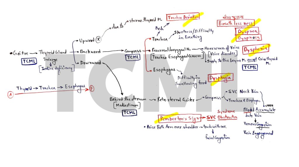
GI bleeding

Overview- 1. Epistaxis 2. Hemoptysis 3. Hematemesis 4. Hematochezia 5. Melena6. Hematouria Blood from Nose (Epistaxis) It is also called Epistaxis. Some important questions about nose bleeding. Question 1. Epistaxis is due to the rupture of Kiesselbach’s plexus which is present at little’s area of anterior inferior part of the nasal septum. Question 2. Four […]

GI bleeding Read More »

Thyroid gland

Overview- 1. Dyspnea 2. Dysphonia 3. Dysphagia 4. Tracheal Deviation 5. Retrosternal goiter 6. Pemberton’s Sign Thyroid gland present in neck region and it have two lobe (Right and left) and both lobe are connect with isthmus. Thyroid gland present in front of the tracha and it secret three hormone 1. Thyroxine or tetra iodo

Thyroid gland Read More »

Brain blood supply

Overview- 1. Vertebral Artery- 4th part (Five branches)2. Basilar Artery- Five branches 3. Internal Carotid Artery (ICA)- 4th part (Five branches)4. Circle of Willis (COW)- Draw hexagon 5. Cerebellum blood supply- Three artery 6. Clinicals- Stroke, Ataxia, PICA syndrome (Wallenberg syndrome or Lateral medullary syndrome) NOTE: Vertebral, and Internal carotid artery- • Both artery are

Brain blood supply Read More »

Femoral artery

Femoral artery is the main artery of lower limb and here we study femoral artery into some points that are given below. Overview- A. Inguinal ligament B. Femoral triangle – Femoral sheath C. Femoral artery branches – Trochanteric and cruciate anastomosis D. Saphenous opening/hiatus – Content E. Clinicals- Femoral artery pulse – Mid inguinal point

Femoral artery Read More »

Aorta part’s

If we want to understand the blood supply of the whole body, then we have to first read the branches of the aorta because aorta supplies oxygenated blood to our body. Here we will study the main arteries of the body into two parts. 1. Aorta part’s and it’s branches 2. Whole Body Blood Supply-

Aorta part’s Read More »

Pterion

Overview- We covered pterion anatomy into below points. 1. Fontanelle- Pterion2. Location- Temporal fossa3. Formation 4. Artery- Middle meningeal artery 5. Nerve- Auriculotemporal nerve6. Hematoma7. Surgical landmark 8. Fracture 1. In pediatrics we read four fontanelle- A. Anterior fontanelle or BregmaB. Posterior fontanelle or LambdaC. Anterolateral fontanelle or Pterion D. Posterolateral fontanelle or Asterion Mnemonic:

Pterion Read More »

Temporal fossa

Overview- 1. Boundaries- Anterior, posterior, superior, inferior, lateral, and medial. 2. Communication- Infratemporal fossa. 3. Content- Muscle, artery, and nerve. ● It is a fan shape, shallow fossa. ● Two in number and present on anterolateral side of the skull. ● Temporalis muscle (Fan shape muscle) are attached on it. 1. Boundaries- The boundaries of

Temporal fossa Read More »